The NanoSIMS 50L is a secondary ion mass spectrometer (SIMS), manufactured by CAMECA SAS. The instrument is designed for a high spatial resolution mass spectrometry, and its beam size can achieve a diameter of ~ 50 nm under optimum settings. Virtually all the elements in the periodic table can be ionised by using one of the two types of primary ion source: Cs+ and duoplasmatron (O−) sources.
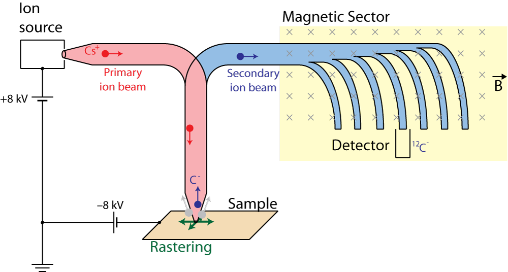
Figure 1: A beam of primary ions, generated in a source, is accelerated and focused to a well-polished sample at normal incidence. This ionises and ejects a fraction of the particles that reside near the surface. The ejected ions are called secondary ions, and these are transferred to the magnetic sector where they are separated due to mass-to-charge ratio. The magnetic sector of the 50L Model has a radius of 650 mm and consists of seven detectors. This enables a multi-collection of up to seven distinct ion species across a wide mass range (factor of ~ 22 from the lowest mass to highest). Each detector is installed with an electron multiplier (EM) and a Faraday cup (FC). EMs and FCs are interchangeably used depending up on the type of analysis — in general, EMs are favoured for a type of analysis that uses a low-current primary beam, such as secondary ion imaging, whereas FCs may be preferred for a high-current analysis, such as high-precision isotope measurement.
Secondary Ion Imaging
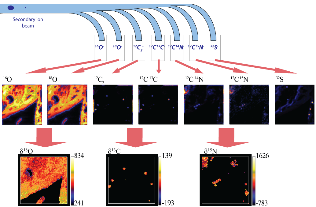
By rastering the primary ion beam (i.e. by deflecting the primary ion beam over the sample surface), the NanoSIMS can perform a pixel-by-pixel collection of secondary ions — "secondary ion imaging". Owing to the finely focused beam and the seven detectors, the NanoSIMS is, therefore, capable of visualising the elemental/isotopic composition of the sample surface in the microscopic scale, as shown in Figure 2.
Figure 2: In the present example, a primary Cs+ beam was rastered over a surface area of 20 × 20 μm (256 × 256 px). Negatively charged secondary ions of 16O, 18O, 12C2, 12C13C, 12C14N, 12C15N and 32S were separated at the magnetic sector and collected by seven the individual EMs, presenting the secondary ion images of these ion species. Processing the 16O and 18O images, the 12C2 and 12C13C images and the 12C14N and 12C15N images would then, demonstrate the isotopic composition of the sample in oxygen, carbon and nitrogen, respectively.